Soft Tissue Infections
A practical interactive module for UK healthcare professionals, covering three time-critical soft tissue infections — necrotising fasciitis, pyogenic flexor tenosynovitis, and bite wounds — with local West Suffolk Hospital antibiotic guidelines throughout.
Time-critical: Necrotising fasciitis is a surgical emergency. Delay to surgical debridement beyond 6 hours is associated with significantly increased mortality.
What is necrotising fasciitis?
Necrotising fasciitis is a rare but rapidly progressive skin and soft tissue infection characterised by invasion of the deep fascial layers, causing diffuse microvascular occlusion and necrosis. It spreads along fascial planes, often far beyond the visible skin changes, and carries a high mortality if not recognised and treated promptly.
~500
cases per year in the UK.
≤40%
overall mortality — up to 45% with septic shock.
80%
of cases extend from a skin lesion — but the breach may have healed.
>32%
mortality for Type II (Group A Streptococcus) disease.
From “hospital gangrene” to a modern misnomer
An army surgeon is first credited with describing the condition, then known as “hospital gangrene.” Its incidence has risen over recent decades. In the 1990s the media coined the term “flesh-eating bacteria” — a misnomer, since the bacteria do not eat tissue but destroy it through toxin-mediated ischaemia. There is also a common misconception that NF only affects high-risk patients.

Why the layers matter
From superficial to deep, the soft tissues are arranged as the epidermis and dermis (the skin), the subcutaneous fat, the superficial and deep fascia, and finally the underlying muscle. The naming of necrotising soft tissue infections reflects the layer chiefly involved — necrotising cellulitis (dermis/subcutis), necrotising fasciitis (fascia), and myonecrosis (muscle).
In necrotising fasciitis, infection tracks along the relatively avascular fascial planes, which offer little resistance to spread. The fascia’s poor blood supply both accelerates the destruction and limits antibiotic delivery, so the infection advances rapidly and far beyond the visible skin changes. Crucially, the overlying skin can look deceptively normal early on because the disease is destroying the deeper fascial layer first — a key reason the diagnosis is so easily missed.

Absence of pus
A notable absence of frank pus. Instead, a thin, foul, greyish “dishwater” exudate is characteristic at exploration.
Widespread necrosis
Destruction extends from dermis to deep muscle and tendon, accompanied by severe, disproportionate pain.
Rapid progression
Progresses rapidly — potentially to septic shock and death within hours of presentation.
Often trivial injury
Arises from minor skin injuries, muscle strains, insect bites, or post-operative obstetric/gynaecological procedures.
NF occurs in both immunocompromised and previously well, immunocompetent hosts. The approach to diagnosis and treatment is the same in either group.
How tissue is destroyed
A breach in the skin or mucosa allows organisms to enter the soft tissues. A deep tissue injury combined with bacteraemia can seed infection. As part of the repair response, inflammatory mediators draw organisms to the site of injury. The organisms release exotoxins that damage local tissues and occlude capillaries, disrupting the vascular supply. Venous occlusion and microvascular thrombosis cause progressive ischaemia and necrosis, producing the erythema, swelling, and ecchymosis/bullae seen clinically.

Susceptible host
Diabetes mellitus, immunocompromise, excessive alcohol intake, obesity, the sedentary/elderly, and malnourishment.
Portal of entry
IV drug use, hypodermic therapeutic injections, insect bites, skin abrasions, ulcers/skin conditions, and abdominal or perineal surgery.
Why classify?
NF is classified microbiologically into four types. While the type guides antibiotic choice and identifies typical host groups, all types require urgent surgical evaluation. Delay greater than 6 hours is associated with increased mortality regardless of type.
Polymicrobial. The most common type. Often elderly, diabetic patients, pressure ulcers, and post-operative abdominal or perineal infections.
Group A beta-haemolytic Streptococcus (and Staphylococcus aureus). Can affect all ages, often with no underlying disease.
Marine-related organisms and Clostridia infections, which can lead to gas gangrene.
Fungal. Predominantly affects immunocompromised patients.
Type II — the one not to miss in the well patient
- Affects all ages, often with no underlying disease
- No portal of entry found in around 50% of cases
- Bacteraemia is common — frequently presents with rigors
- 30–70% mortality; patients may die within 12 hours without surgical intervention
- Exotoxin-mediated toxic shock syndrome
- More than 25% require amputation
Extremities (~75%)
The limbs are by far the most frequent site of necrotising soft tissue infection.

Neck
Includes Ludwig’s angina and Lemierre’s syndrome.

Fournier’s gangrene
Necrotising infection of the perineum and external genitalia.
Abdominal wall
May involve the abdominal wall, with colonic or peritoneal sources.
Systemic features
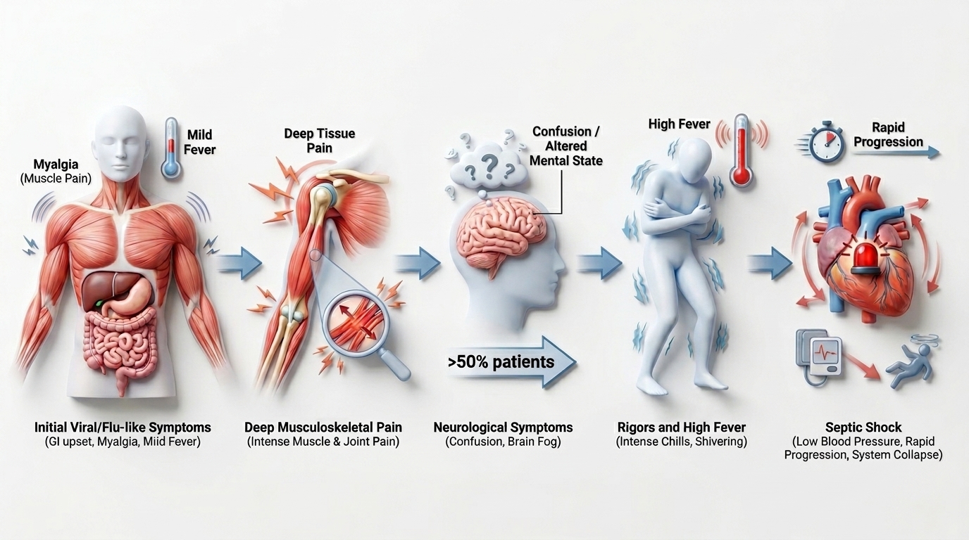
Initial viral/flu-like symptoms (myalgia, GI upset); severe pain deep in muscle/joint; confusion or altered mental state in >50% of patients; rigors and high fever; rapid progression to septic shock.
Local features
Pain out of proportion to examination findings; erythema, tenderness, and warmth; rapid-onset skin rash and blisters; bullae, crepitus, and anaesthesia. The site of entry is often not found.
Activity complete
| Feature | Cellulitis | Necrotising fasciitis |
|---|---|---|
| Threat to life | Not usually life-threatening | Life-threatening |
| Progression | Slow, indolent | Rapidly progressive |
| Pain | Pain not severe | Severe pain, often beyond the visibly involved area |
| Systemic state | No signs of systemic shock | Septic |
| Antibiotic response | Responds to broad-spectrum antibiotics | Does not respond to antibiotics alone |
| Margins | Distinct margins | Indistinct margins |
| Skin changes | — | Bullae formation, skin sloughing, tissue necrosis |
Cellulitis can progress to necrotising fasciitis. Reassess any “cellulitis” that deteriorates rapidly, fails to respond, or develops disproportionate pain.
Features that should raise immediate concern
- Pain out of all proportion to clinical findings
- Altered mental state
- Erythema progressing rapidly along the limb
- Fluctuance
- Haemorrhagic bullae
- Skin necrosis
Sepsis with organ hypoperfusion — septic shock
- Hypotension < 90 mmHg systolic
- Hypoxia
- Lactate > 2
- AKI / raised creatinine
- DIC / thrombocytopenia
- Liver dysfunction
Erythema & pain
Tenderness beyond the apparent margin, warmth, and erythema, with pain out of proportion to the visible findings.
Blistering
Blister and bulla formation, with skin changes evolving over hours.
Haemorrhagic bullae, necrosis & crepitus
Haemorrhagic bullae, skin necrosis/sloughing, anaesthesia of the overlying skin, and crepitus are late signs — surgery should not wait for them.

Erythema & pain

Blistering

Haemorrhagic bullae, necrosis & crepitus
NF is primarily a clinical and surgical diagnosis. If clinical suspicion is high, do not delay surgical exploration for imaging or scores.
History & examination
Take a focused history and examine the limb. Mark the area of erythema and record the date and time to track rapid progression.
Sepsis Six
Initiate the Sepsis Six promptly — oxygen, blood cultures, IV antibiotics, IV fluids, lactate, and urine output monitoring.
Bloods & imaging
- Labs: CRP, procalcitonin (PCT), and group & save (G&S), alongside FBC, U&Es, lactate, and clotting
- Imaging: reserve for cases of lower clinical suspicion — it must not delay theatre when suspicion is high
- Point-of-care ultrasound may show a thickened fascial layer and fluid tracking along fascial planes
Bedside / intra-operative finger test
Under local or general anaesthesia, a small incision is made down to the deep fascia and a gloved finger is used to probe along the fascial plane. Findings suggestive of NF include:
- “Dishwater” fluid
- Lack of bleeding
- Greyish necrotic tissue
- Tissues that peel off the fascia with minimal resistance
Viewer discretion is advised. The following video contains graphic clinical and surgical footage of necrotising fasciitis.

Laboratory Risk Indicator for Necrotising Fasciitis (LRINEC)
The LRINEC score combines routine laboratory values to stratify the risk of NF. It is an adjunct only and does not replace clinical judgement.
| Parameter | Value | Points |
|---|---|---|
| CRP (mg/L) | < 150 / ≥ 150 | 0 / 4 |
| White cell count (×10&sup9;/L) | < 15 / 15–25 / > 25 | 0 / 1 / 2 |
| Haemoglobin (g/dL) | > 13.5 / 11–13.5 / < 11 | 0 / 1 / 2 |
| Sodium (mmol/L) | ≥ 135 / < 135 | 0 / 2 |
| Creatinine (µmol/L) | ≤ 141 / > 141 | 0 / 2 |
| Glucose (mmol/L) | ≤ 10 / > 10 | 0 / 1 |
A LRINEC score ≥ 6 is a reasonable cut-off to rule in NF, but a score < 6 does not rule out the diagnosis. There are no prospective trials validating the LRINEC score.
Principles of management
Survival depends on early diagnosis and emergency surgery combined with aggressive resuscitation. This is a multidisciplinary emergency from the outset.
Recognise & escalate
Early diagnosis and emergency surgical referral. Immediate multidisciplinary input — surgeons, intensive care, and microbiology.
Broad-spectrum antibiotics & resuscitation
Commence broad-spectrum antibiotics and aggressive resuscitation, with supportive care in intensive care.
Surgical debridement
Serial debridement until no further necrosis or infection is seen — removing all necrotic skin and tissue, and any tissue of questionable viability.
Constant re-evaluation
At least a 10 mm margin of healthy fascia, with sampling from healthy fascia and planned re-look procedures to confirm source control.
Trust Microbiology Guideline · CG10239
ALL cases must be discussed urgently with the duty Consultant Microbiologist. Doses are for adults — use the Children’s BNF for paediatrics. Antibiotics never replace surgery.
| Indication | Regimen |
|---|---|
| Core regimen | Meropenem 2 g tds IV AND Clindamycin 1.2–2.4 g qds IV |
| Consider adding | Linezolid 600 mg bd IV — if MRSA colonised / at risk, IVDU, or recurrent skin & soft-tissue infection |
| Consider adding | Gentamicin 5 mg/kg ideal body weight |
| Marine / seafood exposure | Ciprofloxacin 400 mg bd IV (covers Vibrio) |
Penicillin allergy: clindamycin, linezolid, gentamicin and ciprofloxacin are safe. Meropenem and other beta-lactams may be given cautiously if the previous reaction was a rash but not anaphylaxis. Review the clindamycin dose by 48 h post-op. IV immunoglobulin (2 g/kg) is considered case-by-case for strep/staph NF when other measures fail.
🔪 Why clindamycin?
Clindamycin is a protein-synthesis inhibitor — it switches off toxin (superantigen) production by Group A Streptococcus, independent of bacterial load. That is its role beyond simple bacterial killing.
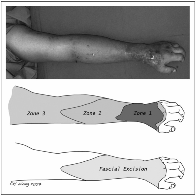
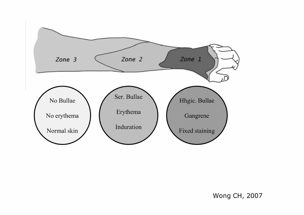
Necrotic tissue
Completely excised, up to the muscle layer.
Infected but salvageable
Meticulously assessed. Fascia must be excised with at least a 10 mm margin of healthy fascia.
Non-infected skin
Left alone. However, any area of skin and subcutaneous tissue that can be easily lifted off the muscle is involved and must be excised.
🩹 Plastic surgery input
Early involvement for soft tissue reconstruction once source control is achieved.
🌀 VAC therapy
Negative-pressure wound therapy to manage large wounds and bridge to definitive cover.
🧑⚕️ Tissue viability
Specialist nursing for complex wound care and dressing strategy.
🦵 Physiotherapy
Rehabilitation, including following amputation where limb salvage is not possible.
⚠️ Why NF is so often missed
- Early diagnosis is notoriously difficult and misdiagnosis is common
- Symptoms mimic other infections — frequently labelled “cellulitis”
- Delay in surgical exploration through failure to recognise the signs and lab findings
- Over-reliance on imaging
- The consequence of delay is loss of limb and loss of life
In one study, NF was initially misdiagnosed 71.4% of the time. Maintain a high index of suspicion.
32M, intravenous drug user
Presents at 06:00h with a 1-day history of a painful left lower leg, with fever and confusion over the last 3–4 hours. On examination there is erythema and a tender, swollen left leg.
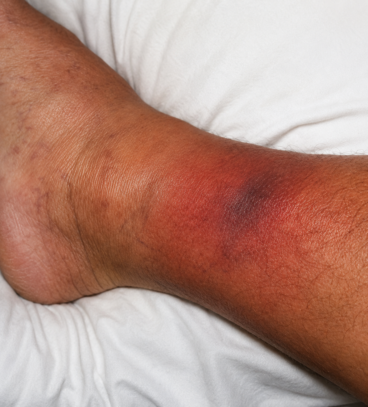
Question 1 — What are your differential diagnoses?
DVT, cellulitis, critical limb ischaemia — and necrotising fasciitis. The combination of disproportionate pain, fever, confusion, hypotension, and a markedly raised CRP/lactate in an IV drug user should raise strong suspicion of NF.
Question 2 — What is the LRINEC score, and how do you interpret it?
CRP 309 (4) + WCC 15.7 (1) + Na 132 (2) + glucose 16 (1) = 8 (Hb and creatinine score 0). A score ≥ 6 supports the diagnosis and ≥ 8 is strongly predictive of NF — but remember a low score would not exclude it, and clinical suspicion already mandates surgical review.
Question 3 — What is your immediate management?
Sepsis Six and aggressive resuscitation, broad-spectrum IV antibiotics, and immediate referral for emergency surgical exploration with intensive care and microbiology input. Mark the erythema with the time. This patient was admitted to ICU and taken to theatre for surgical debridement.
Question 4 — What are the key surgical principles at debridement?
Excise all necrotic and questionable tissue to a margin of at least 10 mm of healthy fascia, sample healthy fascia for microbiology, and plan serial re-look debridements until no further necrosis is seen.
What is it?
Infection within the closed synovial sheath surrounding a flexor tendon — usually 2–5 days after a penetrating injury (rarely haematogenous). A closed space plus a poor blood supply makes this a true hand emergency. Untreated, it can cause tendon necrosis, adhesions, deep-space spread, tendon rupture, and amputation.

9.4%
of hand infections in one large series.
Washout < 24 h
target to theatre when surgery is indicated.
Only 17% febrile
most patients are not febrile — do not be reassured.
54% all four signs
(Dailiana) — absence of the full set does not exclude it.
Why the anatomy matters
Each finger’s flexor tendons glide within a closed synovial sheath. For the index, middle and ring fingers these digital sheaths are separate and end at the level of the distal palm, so they are not continuous with the deeper spaces of the hand. The thumb and little finger are different: the thumb’s sheath (the radial bursa) and the little finger’s sheath (the ulnar bursa) extend proximally and communicate with the common flexor sheath at the wrist. This arrangement dictates how readily infection can track from a digit into the hand and wrist.

Figure 19.1 Synovial sheaths of the hand. Source: Drake R, Vogl A, Mitchell A. Gray’s Anatomy for Students (2nd edn). Philadelphia: Churchill Livingstone; 2010: 760, Fig 7.98.
Q2 — This index-finger infection: will it spread quickly into the hand?
Not especially. The synovial flexor sheath of the index finger is not continuous with the common synovial sheath of the hand itself, so infection is comparatively contained (refer to Figure 19.1).
Q3a — If the infection had started in the thumb, what structure might be rapidly infected?
The common synovial sheath in the hand, and then the wrist — because the sheath of the thumb (radial bursa) is continuous with it.
Q3b — From which other digit would the same rapid spread occur?
The little finger — its sheath (the ulnar bursa) likewise communicates with the common flexor sheath at the wrist.

Horseshoe abscess — because the radial bursa (thumb) and ulnar bursa (little finger) can communicate at the wrist, infection may track from one to the other, producing a horseshoe-shaped collection.

Fixed flexion of the digit. The finger rests in slight flexion — the position of comfort that minimises sheath tension.
Fusiform (“sausage”) swelling. Uniform swelling along the whole digit — Pang et al found this the most common sign at presentation.
Tenderness along the flexor sheath. Tracks the sheath, not just the wound — often the last sign to appear.
Pain on passive extension. Widely held to be the earliest and most reliable sign — gently extend the finger and watch the face.
Described by Allen Kanavel in 1912 and never formally validated — they remain the best bedside tool because imaging and bloods are non-specific. Which sign would you elicit first? Passive extension — earliest, most sensitive, and least equipment.
Pathway
- Surgical emergency — admit, elevate the hand, IV antibiotics, mark and monitor.
- If caught < 48 h, a trial of IV antibiotics with elevation and close observation may be appropriate.
- Otherwise → urgent irrigation & debridement of the sheath, typically within 24 h.
- Send washout / tissue for culture to guide targeted therapy.
🐟 Case — a puncture from a fish tank
A keen aquarist punctures a finger; three days later all four Kanavel signs are present.
Think atypicals. Water exposure widens the differential beyond Staph/Strep — case reports describe Shewanella and Mycobacterium marinum. Take a detailed exposure history, send appropriate cultures, and flag the water contact to microbiology — empirical cover may need broadening.
Don’t be reassured by an afebrile, well-looking patient. Only ~17% are febrile, and signs are blunted in diabetics, the immunocompromised, and IV drug users. The diagnosis is clinical and time-sensitive — refer early.
There is no dedicated PFT line in the Trust guideline — cover the likely organisms and follow local hand-surgery / microbiology advice, mapping the cause to the right Trust pathway.
| Scenario | Empirical cover |
|---|---|
| Typical (no bite / water) | Cover Staph aureus + streptococci — align with the Trust severe cellulitis / soft-tissue pathway (e.g. IV flucloxacillin ± clindamycin; vancomycin if MRSA). |
| Bite-associated | Use the bite pathway: co-amoxiclav 1.2 g tds IV (covers Pasteurella/Eikenella + anaerobes). |
| Penicillin allergy | Doxycycline + metronidazole; or if IV needed and not anaphylaxis, ceftriaxone 2 g od + metronidazole. |
| Water exposure | Discuss with microbiology — atypical / Gram-negative cover may be required. |
Always discuss with hand surgery and the duty microbiologist, and de-escalate on culture results. Surgery remains the definitive treatment when indicated. The commonest “penetrating injury” behind PFT is a clenched-fist injury or animal bite over a finger — which leads directly into bite wounds.
Scale of the problem
Bites account for about 1% of all ED attendances. Dogs cause the most by volume, but cat and human bites carry a disproportionately high risk of infection.

3–18% infected
crush + tearing injury pattern.
28–80% infected
deep, narrow punctures.
~5 species
median bacterial species cultured per infected bite.
50% / 75%
Pasteurella in dog / cat bites respectively.
Pasteurella canis, Staph, Strep, Capnocytophaga canimorsus, anaerobes. Crush and tearing injury.
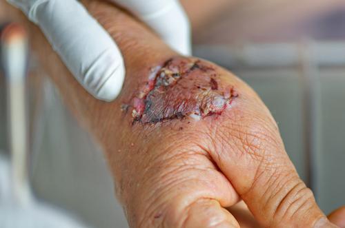
Pasteurella multocida (75%). Deep, narrow punctures inoculate sheaths and joints — high infection risk, may need washout.
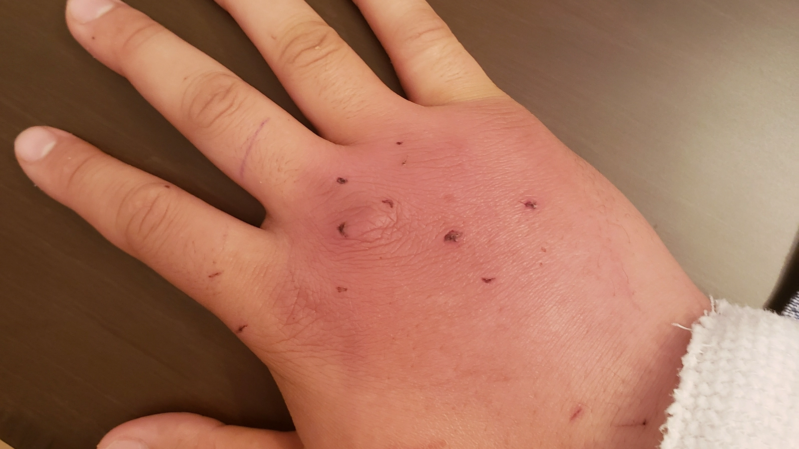
Eikenella corrodens, viridans strep, anaerobes. “Fight bite” over the MCPJ — assume joint/tendon involvement until proven otherwise.
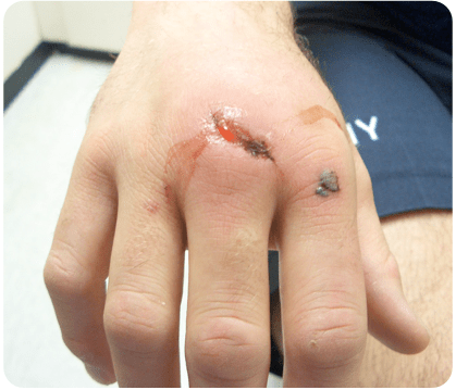
Cat bite, skin broken, no bleeding, healthy adult
Give prophylaxis. For cats the rule is simple — prophylaxis if the bite has broken the skin, even if no blood was drawn. Consider washout if on the hand/wrist.
Dog bite to the hand, deep puncture, drew blood
Offer prophylaxis. Dog bites warrant it if the skin is broken + blood drawn AND a high-risk feature: hand/foot/face/genitals, deep/puncture/crush, contamination, penetration of bone/joint/tendon/vessel, or relevant comorbidity.
Human bite to the knuckle, skin broken
Offer prophylaxis — high-risk area overlying a joint. Examine for joint/tendon involvement (fight bite). Don’t forget the blood-borne virus risk assessment for human bites.
Mosquito bite, itchy, no signs of infection
No antibiotic. Redness/itch can last up to 10 days and rarely gets infected. Watch for erythema migrans (Lyme → NICE NG95) and pain out of proportion (toxin-producers).
🚿 Wound care
Irrigation and debridement as necessary — arguably more important than antibiotics. Deep/contaminated wounds may need surgical exploration.
💉 Tetanus & more
Assess tetanus (booster ± immunoglobulin), blood-borne virus exposure (human bites), and rabies for bites in endemic countries and any bat exposure in the UK.
🧫 Microbiology
Swab if any discharge; send tissue if debrided; send aspirate if aspirated. Reassess if worsening or systemically unwell.
Statistic worth knowing: infected bites grow a median of five organisms, and anaerobes are present in roughly a third — which is exactly why a beta-lactam plus a beta-lactamase inhibitor (co-amoxiclav) is the logical single agent.
Trust Bites Guideline · CG10049
Empirical cover for human & animal bites. Always reassess and de-escalate on culture results.
| Indication | Regimen |
|---|---|
| First line | Co-amoxiclav 625 mg tds PO OR co-amoxiclav 1.2 g tds IV |
| Penicillin allergy | Doxycycline 100 mg bd PO AND metronidazole 400 mg tds PO |
| Pen-allergy, IV needed, NOT anaphylaxis | Ceftriaxone 2 g od IV AND metronidazole 400 mg tds PO |
| Duration | Prophylaxis: 3 days · Treatment (established infection): 5–7 days |
Doxycycline: avoid concurrent calcium/iron supplements; take with food, sitting or standing. Insect bites: no antibiotic unless infected. Wild/exotic animal bites → seek advice from the duty Consultant Microbiologist.
🐾 Why co-amoxiclav fits perfectly
It covers Pasteurella, Eikenella, staph, strep and anaerobes in one agent — the precise polymicrobial spread you culture from a bite.

🏆 Module Complete
You have completed the Soft Tissue Infections module, covering necrotising fasciitis (recognition, pathophysiology, classification, time-critical resuscitation, and emergency surgical management), pyogenic flexor tenosynovitis, and bite wounds — including the local West Suffolk Hospital antibiotic guidelines.
Confirmation of completion
Necrotising fasciitis · Flexor tenosynovitis · Bite wounds